3D Printed Tumor Models Enhance Surgery Precision
At Southampton University Hospital, surgeons are turning to the power of 3D printing to craft highly detailed tumor models, generated from CT and MRI scans. This initiative aims to enhance the precision of intricate cancer surgeries, focusing mainly on patients suffering from hilar cholangiocarcinoma—a complex, challenging form of bile duct cancer. This mirrors recent trends of leveraging 3D printing within the medical industry, as an increasing number of healthcare professionals are recognizing its transformative potential in improving patient wellbeing.
Hilar cholangiocarcinoma, a cancer that starts in the bile ducts connecting the liver to the gallbladder, presents a unique set of obstacles for medical professionals. Although surgeons have access to CT and MRI scanning to help determine the severity of the tumor, the lack of clear preoperative visibility can result in significant invasive measures being taken.
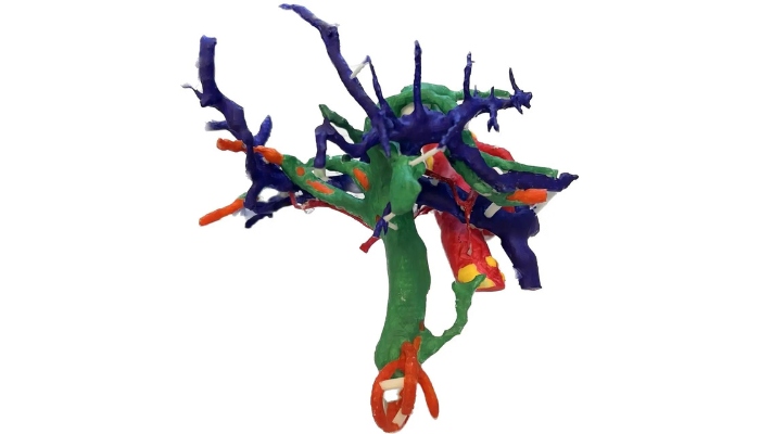
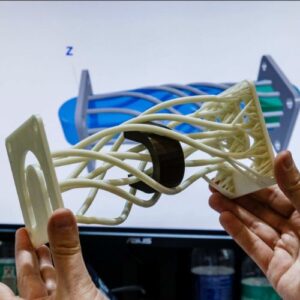
3D printed liver model used to aid in preparation and planning of tumor removal.
Indeed, due to the complicated nature of this cancer, surgeons frequently encounter unforeseen challenges during surgery for which they couldn’t adequately prepare for beforehand. This in turn makes it difficult for surgeons to determine the feasibility of a complete tumor removal until the surgery is already well underway. This can lead to irreversible decisions during the operation with long-term adverse outcomes for patients. Enter 3D printing.
Improving Cancer Treatment With 3D Printing
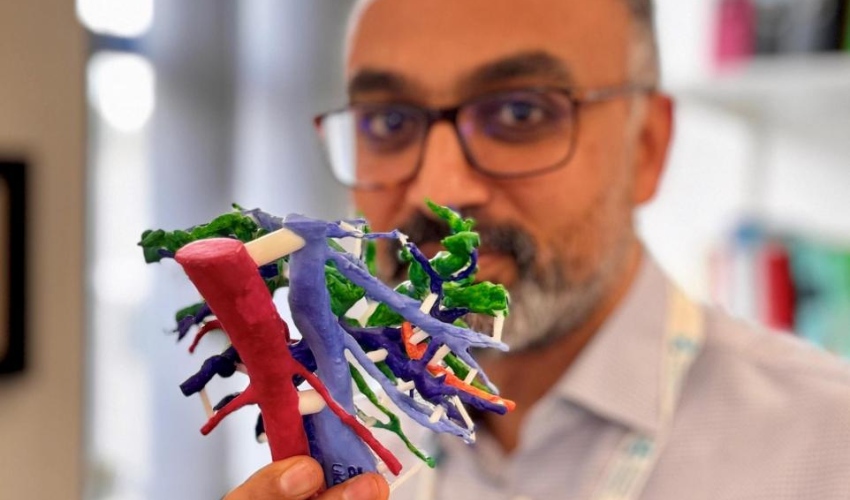
This project, led by Mr. Arjun Takhar, a consultant hepatobiliary and pancreatic cancer surgeon, seeks to directly improve outcomes by converting patient-specific data into detailed 3D models. The goal? To allow surgeons to assess the tumor and its intricate connections. His team, supported by the PLANETS Cancer Charity, uses 3D printing technology to transform CT and MRI scan data into realistic, patient-specific models.
These 3D printed replicas offer a tangible, scaled representation of the patient’s tumor, enabling surgeons to more thoroughly plan and make informed decisions before stepping into the operating room. By offering the ability to provide a clearer understanding of a patient’s surrounding anatomy, such as blood vessels, bile ducts, and other nearby structures, surgeons can now proactively reduce the risk of invasive missteps during surgery. Mr. Takhar expressed optimism about the broader impact of incorporating 3D printing in combating this cancer when stating,
There is also an added advantage that a 3D model would also help in teaching trainee surgeons the nuances of liver anatomy in relation to these complex tumors. This is a unique opportunity to use a novel technology to help patients with a difficult disease, and we foresee adoption of the technology in patients with other liver tumors, too.”
Southampton University Hospital’s use of 3D printing in cancer surgeries marks a notable advancement in medical technology. As 3D printing gains traction in healthcare, these developments have the potential to reshape surgical approaches, offering improved outcomes for patients dealing with complex conditions like hilar cholangiocarcinoma and other cancers. To learn more about Southampton University Hospital’s use of 3D printing in surgery, click here.
What do you think about the potential impact of Southampton University Hospital’s use of these 3D printed tumor models? Let us know in a comment below or on our LinkedIn, Facebook, and Twitter pages! Don’t forget to sign up for our free weekly newsletter here, the latest 3D printing news straight to your inbox! You can also find all our videos on our YouTube channel.
*All Photo Credits: PLANETS Cancer Charity