Researchers 3D print functioning human heart pump
At the University of Minnesota, researchers have 3D printed a functioning centimeter-scale human heart pump. Scientists believe that this discovery could have some major implications for studying heart disease, one of the leading causes of death in the United-States, killing about 600,000 people per year. Bioprinting techniques have developed tremendously in recent years, and research teams around the world are studying how such techniques could be used to cure heart conditions. The ultimate goal being to reproduce a human heart that could be transplanted into patients. Even though experts estimate that we will need to wait between 10 to 15 years for this to be possible, breakthroughs such as this one at the University of Minnesota indicate that other solutions exist beyond bioprinting organs.
More precisely, the researchers had tried in the past to 3D print heart muscle cells that were derived from pluripotent human stem cells, in other words cells with the potential to develop into any type of cell in the body. Researchers aimed to reprogram these cells to heart muscle cells and then use specialized 3D printers to print them. The problem was that scientists could never reach critical cell density for the heart muscle cells to actually function.

A team of researchers from Tel-Aviv University (TAU) successfully 3D printed a heart using human cells back in April 2019. Researchers estimate that it will take an additional 10 to 15 years before this solution is viable.
Therefore, researchers at the University of Minnesota flipped the process. Brenda Ogle, lead researcher on the study explains: “With our team’s expertise in stem cell research and 3D printing, we decided to try a new approach. We optimized the specialized ink made from extracellular matrix proteins, combined the ink with human stem cells and used the ink-plus-cells to 3D print the chambered structure. The stem cells were expanded to high cell densities in the structure first, and then we differentiated them to the heart muscle cells.”
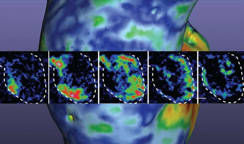
As a result, for the first time ever, the team was able to achieve the goal of high cell density within less than a month to allow the cells to beat together, just like a human heart. Ogle’s team explains that this discovery is a critical advance in heart research because it shows how they were able to 3D print heart muscle cells in a way that the cells could organize and work together. Given that the cells were differentiating right next to each other, this process was more similar to how the stem cells would grow in the body and then undergo specification to heart muscle cells. With these structures, “we can introduce disease and damage into the model and then study the effects of medicines and other therapeutics,” she explains.
Today, this model measures about 1.5 centimeters and will need to undergo multiple tests. In fact, it was specifically designed to fit into the abdominal cavity of a mouse for further study. “All of this seems like a simple concept, but how you achieve this is quite complex. We see the potential and think that our new discovery could have a transformative effect on heart research,” Ogle concludes. You can find more information HERE.
What do you think of this latest breakthrough? Let us know in a comment below or on our Facebook and Twitter pages! And remember to sign up for our free weekly Newsletter, to get all the latest news in 3D printing send straight to your inbox!