Veterinary 3D Printing Helps in Dog’s Hip Surgery
Whether on two legs or four, and regardless of fur, feathers or hair, advances in medical 3D printing have been benefitting humans and animals alike. The veterinary world in particular has seen many 3D printed prosthetic limbs and parts with can easily be attached. However, in both cases, the use of 3D printing has been instrumental in allowing doctors and surgeons to gain more information and preparation before operating by printing replica organs that give more tangible familiarity with the necessary job before reaching the operating table. Such was the case recently at Texas A&M where veterinarians performed a double hip replacement surgery and were able to successfully carry out the delicate procedure thanks to a 3D-modeled and printed replica of the canine’s hip joints.
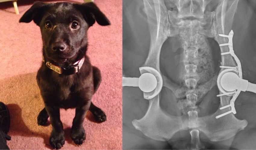
The recipient of the 3D surgical prep was Ava, a Labrador Retriever with a history of hip troubles that goes back 10 years. After realizing that she didn’t ever jump, Ava’s owners decided to get her x-rayed whereupon vets discovered that her hips were not correctly in their sockets. She underwent hip replacement surgery for both hips in 2013 and 2014, and was able to enjoy normal range of motion until veterinarians again discovered an issue in 2020. “Over many years, the artificial ball had worn away the plastic liner protecting the metal wall of the artificial joint,” explains Dr. Brian Saunders which again caused a dislocation and added further complication with metal debris from the old prosthetics building up into a granuloma, which can block off the hip and potentially cause damage internally.
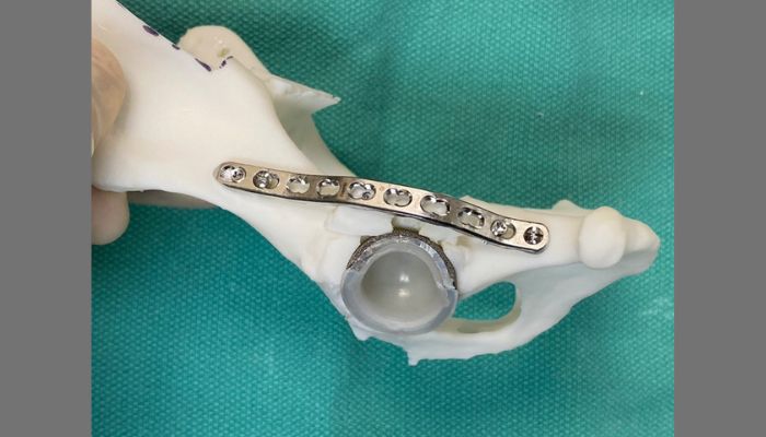
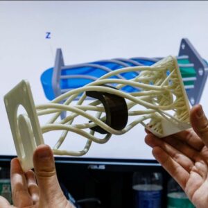
The 3D printed model of the hip and implant.
No question, then, that surgery had to be performed to swap the worn out hip replacements and remove the granuloma before Ava was more negatively impacted. In order to make it happen, Dr. Saunders and his team were able to utilize more cutting-edge technology with the help of 3D scanning and printing. First, a highly precise model of Ava’s hip joints was created through CT scanning, after which the bone models were 3D printed into a one-to-one replica which doctors could use to view and plan the surgical process.
“Basically, we printed a replica of Ava’s dislocated hip joint and planned exactly how to perform the revision operation using the 3D bone models. In fact, we sterilized the plastic models and used them in the operating room to help guide the revision surgery.” continues Dr. Saunders, “Planning using 3D models and collaborating with the Soft Tissue team were two huge contributing factors to our success. […] Thankfully, the second hip wasn’t quite as affected as her first and we already had her 3D bone models from the recent surgery, so the second hip revision surgery was more straightforward.”
The end result is a new lease on life for Ava the dog, who is reportedly back to zooming around the backyard thanks to the diligence and planning of the Texas A&M veterinary team. Ada may be 12 years old, but with her new hips, she can play like a puppy for many more years to come. To learn more about Ada and the Texas A&M School of Veterinary Medicine and Biomedical Sciences, click HERE.
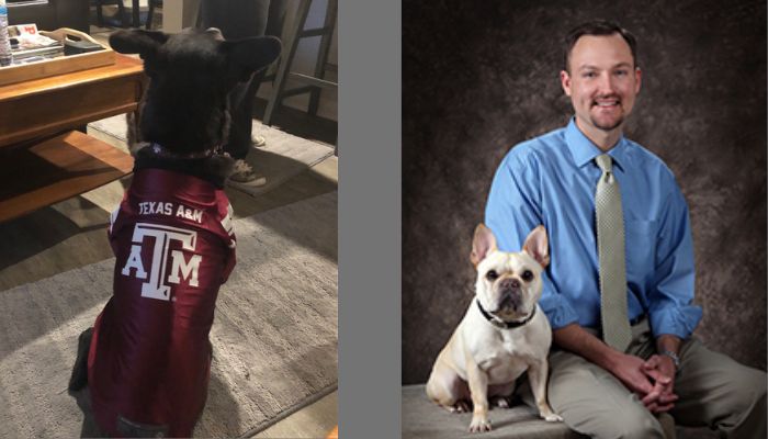
Ava (left), feeling good and sporting Aggie pride after the successful surgeries, and Dr. Brian Saunders (right), service chief of the Texas A&M veterinary hospital.
What do you think about the role of 3D printing in surgery preparation? Let us know in a comment below or on our LinkedIn, Facebook, and Twitter pages! Don’t forget to sign up for our free weekly Newsletter here, the latest 3D printing news straight to your inbox! You can also find all our videos on our YouTube channel.
*All image credits: Texas A&M