





Notre Dame Researchers Develop Method for 3D Printing Blood Capillary Networks
Researchers at the University of Notre Dame have successfully 3D printed blood capillary networks using a surprising technique. Before exploring their method, it’s worth understanding why 3D printing blood capillary networks matters in the first place. In the United States alone, over 100,000 people are waiting for organ transplants, and donor organs remain in short supply. If we could 3D print organs out of a patient’s own cells, it could not only address the shortage, but potentially eliminate the risk of the body rejecting the organ altogether.
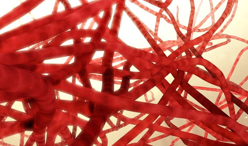
That’s why researchers have been working for decades to 3D print organs. One central challenge, however, is replicating the scale and complexity of the body’s vascular networks, especially capillaries. A blood capillary network is an interlacing web of the body’s smallest blood vessels. These microscopic tubes deliver oxygen and nutrients to cells and remove waste products, making them a necessary component of any bioprinted tissue model.
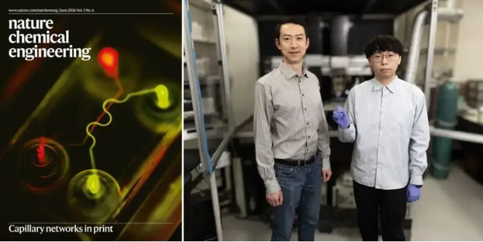
(Left) Capillaries visualized with fluorescent imaging, appeared on the cover of Nature Chemical Engineering. (Right) Yanliang Zhang and Yuxuan Liao, doctoral student and lead author of the study. (Photo by Wes Evard / Notre Dame College of Engineering)
“Printing blood vessels that mimic natural living systems is very difficult since the vessels vary in size,” said Yanliang Zhang, the Advanced Materials and Manufacturing Collegiate Professor in the Department of Aerospace and Mechanical Engineering. “Getting the smallest vessels right, without losing scalability and structural integrity, has remained one of the greatest challenges to current state-of-the-art bioprinting.”
Combining Two 3D Printing Methods
To print vascular networks containing capillaries fewer than 10 micrometers in diameter (smaller than the finest human hair!), the team use two techniques, each responsible for separate components. First, the matrix, a soft, gel-like scaffold that mimics real tissue, is printed by extrusion, which uses pressure to dispense biomaterials one layer at a time. Once a section of the matrix is printed, thin threads of gelatin are deposited within it by aerosol jet printing (AJP), which are later removed to leave behind channels within the matrix. AJP achieves aerodynamic focusing using a sheath flow, which allows the channel size to be adjusted with precision. The range is significant: the gelatin threads can span from hundreds of micrometers down to just a few, mimicking the varied architecture of natural vessels.
“We’ve reached a major breakthrough in the field of bioprinting,” added Zhang. “Achieving capillary-scale resolution in bioprinting is an important step towards engineering fully functional tissues and complete organs.”
Machine Learning for Optimal Printing Parameters
The team also incorporated machine learning to autonomously optimize printing parameters for each desired vascular configuration. The machine learning framework they developed let them fine-tune ink flow and sheath gas flow rate, identifying the ideal combination of printing parameters for a given channel size.
“Machine learning is a very powerful tool that helps us to identify the optimal parameters much more quickly compared with the conventional trial-and-error method,” Zhang said. “Incorporating autonomous optimization has yielded a large boost in efficiency for realizing the desired quality and precision of the printed channels.”
Using this dual-printing approach alongside machine learning, the researchers successfully fabricated hierarchical vascular networks in one, two and three dimensions. Certain channels were then seeded with endothelial cells, which form a thin, single-layer lining called the endothelium on the inside of all blood vessels, heart chambers, and lymph vessels. Adding these endothelial cells led to the formation of single-cell layers akin to those seen in living human tissue.
“We are very pleased to see living cells rapidly attach and spread along the inner walls of the channels, ultimately replicating the barrier function of real human capillaries,” Zhang said. “It’s a big step forward in showing that our hybrid-printed networks are capable of supporting living tissue structures without leakage.”
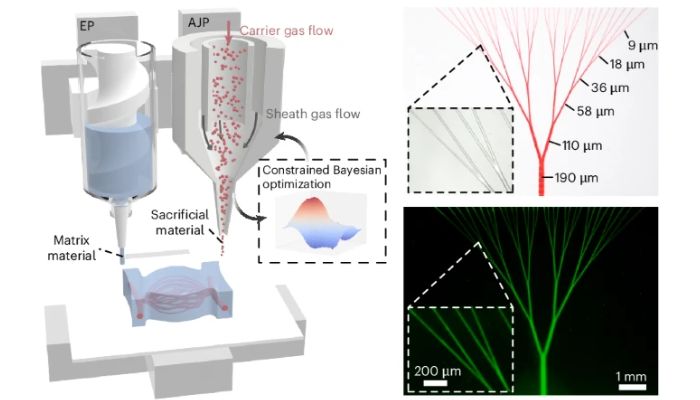
The tissue-mimicking matrix is printed via extrusion (left), while the gelatin-based sacrificial material is deposited via aerosol jet printing (right). Once printing is complete, the system is immersed in warm water, and the gelatin-based material, now liquified, is removed, leaving behind precise channels within the matrix material (far right). (Image Credit: Yuxuan Liao, et al.)
Potential Applications for 3D Printed Blood Capillary Networks
If incorporated into biomimetic tissue models, such designs could be used for regenerative medicine and organ engineering applications, as well as “organ-on-a-chip” models to test drugs for safety and efficacy. Additionally, the use of patient-specific cells to form these models could help develop personalized medicine, where therapeutic responses could be evaluated within the model system prior to full-scale treatment.
Looking ahead, Zhang’s ultimate aim, according to Notre Dame, is to develop an autonomous, intelligent bioprinter capable of producing fully functional tissues and organs such as the heart, kidney and liver. In the near future, his research group and their collaborators at Harvard have secured new funding from the National Institutes of Health to design and build an even more powerful version of Zhang’s hybrid bioprinter and pursue the fabrication of lab-grown organs.
This study was supported by the National Institutes of Health, and featured on a recent cover of Nature Chemical Engineering.
What do you think of 3D printing blood capillary networks? Let us know in a comment below or on our LinkedIn and Facebook pages! Don’t forget to sign up for our free weekly Newsletter here, the latest 3D printing news straight to your inbox! You can also find all our videos on our YouTube channel.
*Cover Image: Capillaries. Image Credit: Cleveland Clinic







Capillary networks are incredibly tricky to recreate. @dashmetry
The idea of 3D printing organs to solve the transplant shortage and eliminate rejection is truly a dream. It’s fascinating how they’re tackling the microscopic complexity of capillaries – makes me think about all the intricate systems in our bodies we often take for granted. Speaking of tiny details, I’ve been playing a lot of Heardle lately, and it’s such a fun way to test your song recognition skills starting from just that first little snippet. It’s got that same thrill of recognizing something from just the smallest detail. Do you think there’ll ever be a “Heardle” for scientific breakthroughs, where you guess the field from just the first sentence of a paper?