Wake Forest researchers create lab model of the human body
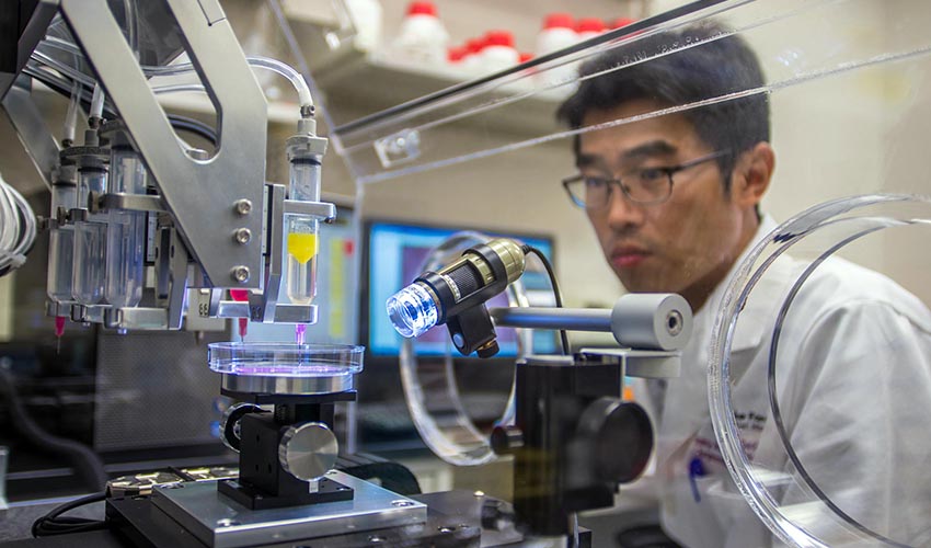
Using bioprinting techniques, researchers at the Wake Forest Institute for Regenerative Medicine have developed a miniature lab model of the human body. This model contains miniaturized human organs such as the heart, lungs and liver, and will be used to detect the adverse effects of drugs before they are prescribed to patients. Additionally, screening potential pharmaceuticals in this way could bring new drugs to market sooner, lower the costs of clinical testing and reduce animal testing.
In the paper published by the journal of Biofabrication, the researchers detail how the miniature organs were created and how the human organ tissue system works. Because of the specified individual requirements of each type of tissue, many biofabrication techniques were employed to create each organ. Tiny samples of human tissue cells were isolated and engineered into miniature versions of the human organs – meaning they can perform the same functions as they do in the human body but at a much smaller scale. Each of miniature organs are essentially tiny 3D tissue-like structures, about one millionth in size of an adult human organ.
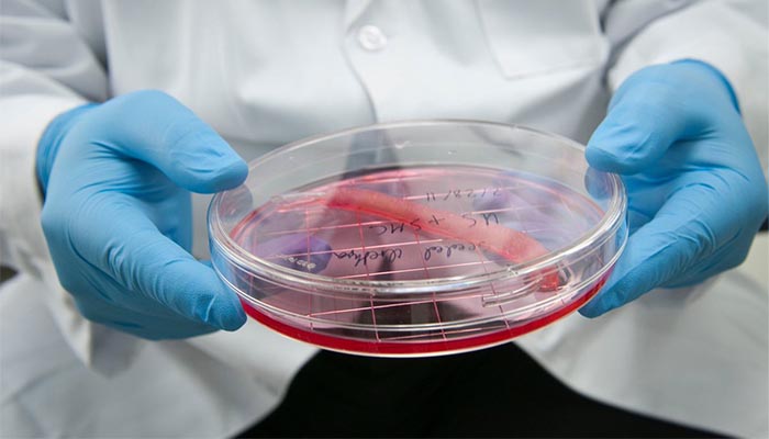
Image via Wake Forest Institute of Regenerative Medicine
In healthcare, 3D printing has been adopted by hospitals and clinics to make precise surgical models that mimic the anatomy of patients. These custom-made anatomical models can prepare surgeons for surgery in a way that was impossible before. Although different, the idea here is also to study the human body and how it will react to a drug. Anthony Atala, Professor at WFIRM commented: “The most important capability of the human organ tissue system is the ability to determine whether or not a drug is toxic to humans very early in development, and its potential use in personalized medicine. Weeding out problematic drugs early in the development or therapy process can literally save billions of dollars and potentially save lives.”
Bioprinting is opening the door to personalized medicine at a quicker rate than ever before. Even though we are still far away from being able to create functional human organs ready for transplantation, there are a number of applications we can already benefit from. In this case, the main objective is to measure the toxicity of drugs before they are prescribed to humans. According to the scientists, many 2D cell cultures or animal testing does not measure correctly the toxicity of a pharmaceutical because the systems are not complex enough or similar enough to humans. Aleks Skardal, PhD, explains: “In order to model the body’s different responses to toxic compounds, we needed to include all of the cell types that produce these responses.”
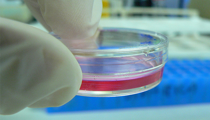
Image via Flickr
As one can imagine, this miniature system was not easy to create. In fact, at WFIRM, researchers have been working over the past 30 years on building a full-scale human organ for transplantation into human patients. Thomas Shupe, PhD of WFIRM concludes: “Creating microscopic human organs for drug testing was a logical extension of our work.”
What do you think of this new research to measure the toxicity of a drug? Let us know in a comment below or on our Facebook and Twitter page! Don’t forget to sign up for our free weekly Newsletter, with all the latest news in 3D printing delivered straight to your inbox!