Improving Cardiovascular Surgical Outcomes with 3D Printed Grafts
What is the most dangerous disease in the world? Although most would point to cancer, it is actually heart disease that takes that role and even curing it can be deadly for sufferers of a variety of cardiovascular conditions. Thankfully, there may be a solution. A research team from Donghua University and Shanghai Jiao Tong University have published a study showing that 3D printed electrospun vascular grafts loaded with TMP (tetramethylpyrazine, a chemical compound found in the Japanese food nattō and fermented cocoa beans), could improve cardiovascular disease treatments by reducing thrombosis (occurring when blood clots block blood vessels and commonly seen due to immobility) and retrain aneurysmal dilation post-surgery.
According to the British Heart foundation, there are around 620 million people living with cardiovascular diseases across the world, an estimated 1 in 13 people. Not only that, but it is the leading cause of death, accounting for at least a third worldwide, while cancers are responsible for about one-in-five deaths. The treatment often includes vessel replacement due to severe stenosis, or total narrowing of arteries in the heart, but current standards still face challenges such as thrombosis. This is where these 3D printed electrospun vascular grafts are expected to make a significant difference.
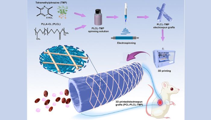
An illustration of the 3D printed electrospun vascular graft filled with TMP (image credits: Burns & Trauma)
Using 3D Printed Vascular Grafts to Treat the Heart
The grafts were made by combining electrospinning, an electrohydrodynamic process to create fibers, and an unspecified 3D printing process. They include an inner layer made of electrospun poly (L-lactic-co-caprolactone) (PLCL) nanofibers and an outer layer of 3D printed poycaprolactone (PCL) microfibers. A dual layer design was chosen for stability and flexibility. TMP, which was derived from the Chinese medicine Ligusticum chuanxiong, was then added for antiplatelet and anticoagulant properties.
The result? In vitro tests showed that the 3D printed grafts were able to reduce platelet adhesion, a necessity when fighting against thrombosis, and they exhibited good compatibility with human cells. Moreover, experiments in rats where abdominal aortas were replaced with the 3D printed grafts showed excellent biocompatibility and mechanical strength over six months. Most importantly, neither acute thrombosis nor significant aneurysmal dilatation were detected, showing their huge potential for improving outcomes in patients who require vascular grafts.
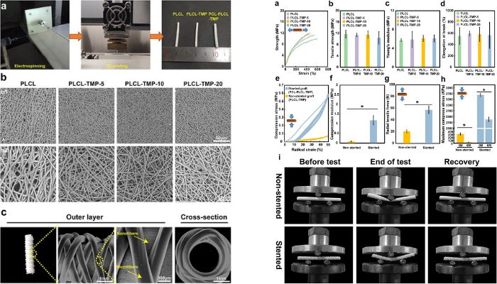
(Left) A closer look at the preparation of the graphs; (right) the results from different tests (image credits: Burns & Trauma)
Dr. Hongbing Gu, a lead researcher, concluded, “This study marks a significant advancement in vascular tissue engineering. The combination of electrospinning and 3D printing, along with the incorporation of TMP, has resulted in a vascular graft that not only meets mechanical requirements but also exhibits excellent blood compatibility. These findings pave the way for future clinical applications.” Future research is expected to focus on large animal models, but in the meantime you can find out more in the published paper HERE.
What do you think ? Let us know in a comment below or on our LinkedIn, Facebook, and Twitter pages! Don’t forget to sign up for our free weekly newsletter here for the latest 3D printing news straight to your inbox! You can also find all our videos on our YouTube channel