Harvard Researchers Develop Microbial Ink to 3D Print Living Materials
What if you were told that 3D printing is getting closer and closer to revolutionizing science? A team of researchers at Harvard University and Brigham and Women’s Hospital is rising to the challenge. Recently, they developed a microbial ink or hydrogel to 3D print living materials. According to the researchers, the innovative material would be able to release drugs or remove chemical contaminants from the environment. This could lead to a revolution in cancer research or advanced applications in outer space. The results of the project were published in the journal Nature Communications in the article, Programmable microbial ink for 3D printing of living materials produced from genetically engineered protein nanofibers by Duraj-Thatte et al., where they explained the development process that they carried out over several years, until they were able to demonstrate the effectiveness and opportunities offered by this technology in science.
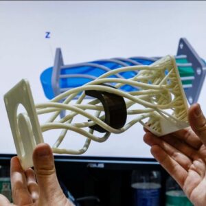
Today we are witnessing a growing synergy between biology and materials engineering, where the implementation of additive manufacturing is also coming into play as a key tool. We have already seen the success story of the University of Colorado, which developed a 3D printable material capable of mimicking complex biological tissues. Or the scientists who created a novel bioink out of ulvan, a type of seaweed, that they believed would promote wound healing. This time, experts from Harvard University created programmable microbial inks from genetically modified protein nanofibers.
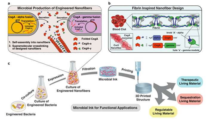
Photo Credits: Harvard University / Brigham and Women’s Hospital
Microbial Ink to 3D Print Living Materials
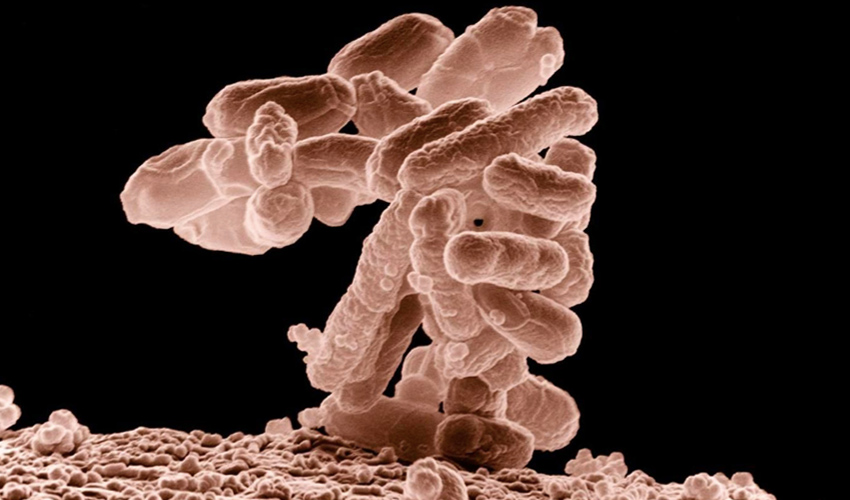
With the idea in mind, the researchers incorporated the bacteria Escherichia coli (also called E.coli) and modified protein nanofibers into the ink. E.coli is a bacterium belonging to the enterobacteria family, which is found in the microbiota of the gastrointestinal tract of homeothermic animals, as well as in humans. A mixture of alginate and E. coli was 3D printed on a calcium chloride surface. This is because the alginate molecules cross-link on this base to form a solidified gel. Regarding the fabrication process itself, in the published article the researchers commented, “Although inkjet printing, contact printing, screen printing, and lithographic techniques have been explored to print microbes, extrusion-based bioprinting has become one of the most widely used techniques due to its simplicity, compatibility with a variety of bioinks, and cost-effective instrumentation.”
Throughout the research process, the experts encountered a number of challenges. One of them concerned the properties of the living materials to be used during the 3D printing process successfully. To this end, the bioinks had to meet two main requirements: a low viscosity for extrusion, but at the same time high enough to retain their shape after printing. For this, the scientists were inspired by the so-called fibrin, a fibrillar protein with the ability to form blood clots through three-dimensional networks.
There is still a long way to go in the development of living materials, although the future is promising. In fact, the Harvard team claims that it could be implemented in research against certain cancers, or even to supply future colonies on Mars. You can find more information about the project in the official publication HERE.
What do you think of the microbial ink from Harvard University and Brigham and Women’s Hospital? Let us know in a comment below or on our Linkedin, Facebook, and Twitter pages! Don’t forget to sign up for our free weekly Newsletter here, the latest 3D printing news straight to your inbox! You can also find all our videos on our YouTube channel.