Basque Scientists Pioneer 3D Printed Blood Vessels for Tissue Engineering
Did you know that in Spain, around 1,144,214 animals are used every year for scientific experimentation? This practice has always been and will always remain controversial. Although animal models are used in medicine to test treatments for diseases, they don’t always replicate human anatomy, for obvious reasons. 3D printing is a technology with the potential to reduce animal experimentation in science in different ways. We’ve already seen this in specific cases in cosmetics and pharmaceuticals. And recently, a study from the research center CIC biomaGUNE in the Basque Country explored the creation of tissue models with artificial blood vessels using 3D bioprinting with nanomaterials.
The researchers behind this breakthrough are Dr. Dorleta Jimenez de Aberasturi, Dr. Uxue Aizarna, and Dr. Malou Henriksen. As part of the Biofunctional Hybrid Materials group at the Basque center, they have tested different techniques to design and create realistic tissue models for scientific research. In particular, they have focused on tissues with artificial blood vessels that mimic the complexity of human arteries and are capable of pulsing in response to external stimuli. How did they do it? What techniques did they use? Keep reading to find out.
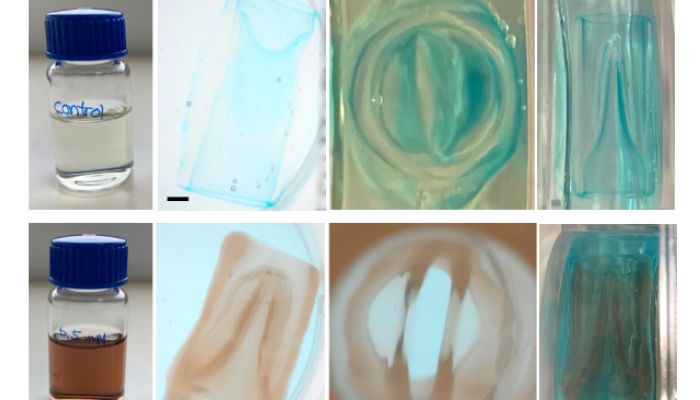
Valves manufactured through volumetric 3D printing. (Credit: American Chemical Society)
One of the secrets behind the breakthrough lies in the “ink” used. The researchers employed methacrylated gelatin as a base, which is cell-compatible, and added gold nanoparticles to it. This combination allowed them to print the structure and control its behavior once printed.
In addition to the ink, there are two standout innovations in this case. On the one hand, the so-called “arteries in the air.” Using an embedded bioprinting technique, in collaboration with Maastricht University, they managed to print soft materials that would normally collapse, creating vessel models with the concentric layers of a real artery. On the other hand, they developed high-speed valves using volumetric 3D printing (together with Utrecht University). This technique projects the shape of an object into the ink and hardens it through light rays. Thanks to this process, they were able to form the entire volume of the structure at once rather than layer by layer, as is the case with most 3D technologies. This made it possible to design and add valves that open and close when activated by an external stimulus.

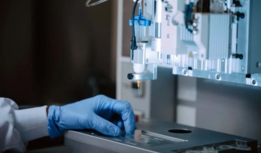
Part of the Biofunctional Hybrid Materials group at the Basque center. (Credit: CIC biomaGUNE)