3D Printed Microfluidic Device to Screen Drugs for Oral Cancer Treatment

Did you know scientists can test how individual cancer patients will respond to drugs before they take them? One method for this research, called functional drug testing (FDT), involves placing patient-derived tumor cells in microfluidic devices to see how they react to different combinations of drugs. Microfluidic devices are meant to simulate the flow of fluids in the body, and the Indian Institute of Technology Hyderabad (IITH) designed novel, 3D printed, microfluidic devices to screen drugs for oral cancer. The goal of this research was to develop a drug screening platform that will allow researchers to see how drugs interact with cancer cells.
IITH designed the 3D printed microfluidic device for the study using Formlabs’ clear resin because they found it to be the best 3D printing material for cultivating the desired cells. Three patients enrolled in the study, whose biopsy samples were used to isolate oral tumor stem-like cells, which were then cultivated further to form spheroids in the microfluidic device. Spheroids are spherical cells, in this case self-aggregating cancer cells, that benefit the research because they help recreate the diverse tumor population and conditions within the body. The result? Spheroids-on-a-chip.

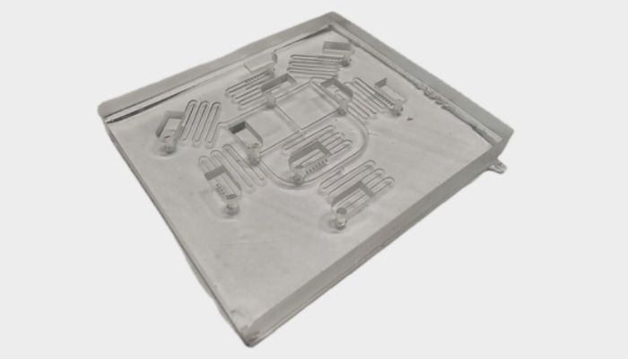
3D printed microfluidic device created by IITH. (photo credit: Mehta, V., Vilikkathala Sudhakaran, S. et al., Journal of Nanobiotechnology, 2024)
This chip, or 3D printed microfluidic device, created by IITH has a two-layer network of serpentine loops for mixing drug combinations, and cylindrical microwells for cultivating the spheroids. The composition allows them to test seven combinations of three well-known drugs for oral cancer: paclitaxel, 5‑fluorouracil, and cisplatin. Because tumors can develop drug resistance, sometimes the best method of treatment involves combining them.
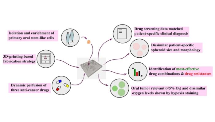
Indeed, the spheroids of patient 1 of the study showed high resistance to all provided drug combinations, while other patients’ spheroids responded positively to certain combinations of drugs or mono-drugs. Therefore, according to the study’s conclusion, the research demonstrates “the influence of tumor differentiation status on treatment responses, which has been rarely carried out in the previous reports.” This means that researchers could determine which drug combinations could be the most beneficial for patients. Additionally, the report stated that “these characteristics also correlated with each patient’s diagnosis from clinical histopathological reports,” further validating the claims.

A graphic representation of the study completed by IITH. (Photo credit: Credit: Mehta, V., Vilikkathala Sudhakaran, S. et al., Journal of Nanobiotechnology, 2024)
The study did not include testing on other types of cells that influence drug responses, or examine how the drug was absorbed by the spheroid upon exposure. Scientists plan to address these limitations with further research and by developing a more complex model. To learn more about IITH’s study, read the original report here.
What do you think of using additive manufacturing to create microfluidic models? Let us know in a comment below or on our LinkedIn, Facebook, and Twitter pages! Don’t forget to sign up for our free weekly newsletter here for the latest 3D printing news straight to your inbox! You can also find all our videos on our YouTube channel.


