Advances in 3D printing for medicine are booming, and researchers at the University of Texas at Dallas have just successfully designed a 3D-printed femur model. This project could provide doctors with a new tool to better prepare bone reconstruction surgeries and develop treatments for bone tumors. The model was developed in collaboration with orthopedic surgeons at UT Southwestern Medical Center, and initial findings from the study have been published in the “Journal of Orthopaedic Research.”
How could this 3D-printed femur facilitate bone reconstruction surgery? The study, focusing on the central part of the bone, defines the precise parameters for producing a 3D femur model suitable for biomechanical testing. Although promising, this technology will still require several stages of research before it can be used in the clinic.
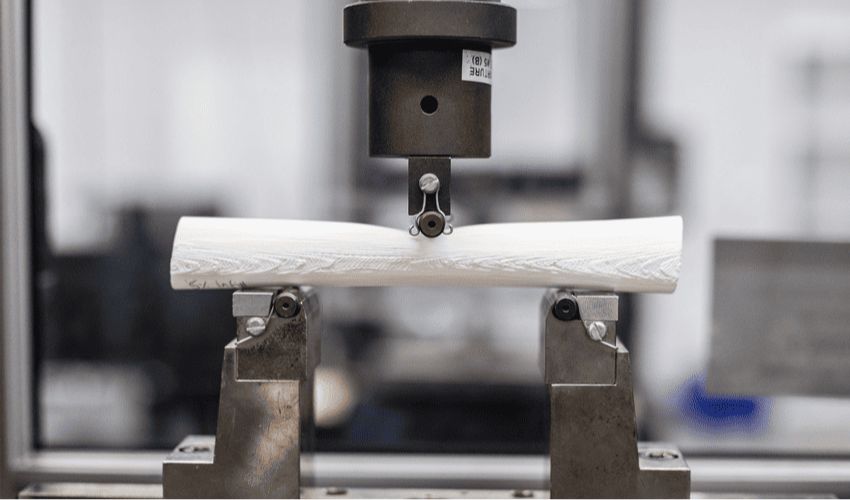
Femur samples made using a 3D printer
A New Approach to Biomechanical Studies with 3D Printing
To examine and evaluate new surgical methods and implants, surgeons carry out biomechanical studies using donor bone or commercially available synthetic bone. This approach enables them to identify the best fixation method and anticipate the bone reaction. However, traditional methods can be costly and time-consuming. In addition, they do not allow solutions to be tailored to specific patient needs, limiting their effectiveness in certain clinical situations.
To address this, researchers at UT Southwestern contacted Dr. Wei Li, a 3D printing specialist at UT Dallas, to work together on a faster, more cost-effective solution for biomechanical studies. “To make plans for surgery, surgeons need to know the geometry of the bone,” says Dr. Wei Li. “With 3D printing, we’re able to print out the femur bone sample with the same geometry of the femur inside the body.”
Kishore Mysore Nagaraja, a PhD student in mechanical engineering at UT Dallas, has improved several versions of the femur through trial and error. Working in the manufacturing laboratory headed by Dr. Wei Li, he carried out various tests on each bone model to assess their mechanical performance. The aim was to make them as close as possible to natural femurs.
Mechanical engineering PhD student Kishore Mysore Nagaraja (left) and Dr Wei Li (right)
The researchers created a bone replica from polylactic acid, an affordable and environmentally friendly polymer that is commonly used in 3D printing. This model represents the central part of the femur and measures almost 20 cm in length and 2.5 cm in diameter. Biomechanical tests have shown that this reproduction offers performance comparable to that of a human femur. What’s more, the production cost of a 3D-printed femur is estimated at around $7.
Dr. Wei Li points out that 3D-printed bones offer a wide range of applications. The polymer used could, for example, replace traditional materials such as titanium in bone repair procedures. He also mentioned the possibility of printing tumors on these bone models to evaluate treatments, or using these replicas to promote the regeneration of human bone tissue.
What do you think of this 3D-printed femur? Let us know in a comment below or on our LinkedIn, Facebook, and Twitter pages! Don’t forget to sign up for our free weekly newsletter here for the latest 3D printing news straight to your inbox! You can also find all our videos on our YouTube channel.
*All Photo Credits: The University of Texas at Dallas