Using Cancer Cells And Computer Modeling, Researchers Have Optimized 3D Bioprinting
Researchers in a multi-university collaboration between the University of Waterloo, Canada, University of Swansea, and University of St Andrews, both UK, have conducted experiments on 3D bioprinted cancer cells over a period of 11 days, to compare to results from a cellular automata model. Their results showed that the model successfully simulated the bioprinted structure; this 3D bioprinting optimization could result in time and monetary savings for future experiments.
The in-vitro experimental method used MDA-MB-231, an aggressive breast cancer cell, which was printed in hydrogel of bioink composed of a mixture of gelatin and alginate. The choice of this bioink is due to the similarity of the two chemicals to native ECM. They monitored the viability of the cellular structure, finding a viability rate of 96 ± 2% after 11 days. The survival of the majority of the cells indicated that the structure was sufficiently porous for glucose and oxygen to permeate for respiration.
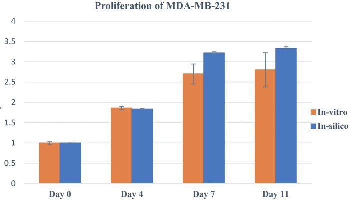
Graph to show the proliferation of the experimental cells in the mathematical model compared to the experimental results. The Y axis represents the number of proliferating cells. (Photo credit: Scientific Reports (Sci Rep))
In-silico refers to experiments which use or integrate computer models (in-silico being pseudo-Latin to mean the silicon chips in computers). For this research, the scientists used cellular automata modeling (modeling using a regular grid of cells), in which most of the parameters were picked from experimental data. A comprehensive in-vitro assay was also done to improve the reliability of the model; that is, the cells were examined to determine their composition to indicate and improve the quality of the model. The final results suggested that the 3D bioprinting optimization using modeling can be used to design experiments to achieve results without needing to conduct assays. The simulated data consistently replicated experimental results for viability and proliferation.
3D bioprinting is an emerging field with important applications in regenerative medicine and tissue engineering. We can look, for example, at its application to create eye tissues, and the Ugandan government recently launched a 3D bioprinter into space in order to conduct tissue experiments in zero gravity. This current experiment to optimize 3D bioprinting by developing a computer model to compare with experimental results is important as it allows the prediction of cellular activities and keeps experimental evaluations to a minimum, saving time and money. For more information, you can find the scientific paper HERE.

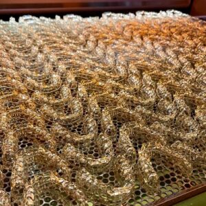
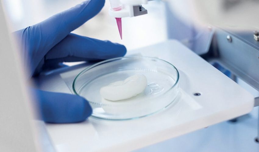
Bioprinting can be used for medical and botanical applications, including this 3D-printed mix of algae and cellulose from the Delft University of Technology (TU Delft). (Photo credit: TU Delft)
What do you think of this 3D bioprinting experiment? Let us know in a comment below or on our Linkedin, Facebook, and Twitter pages! Don’t forget to sign up for our free weekly Newsletter here, the latest 3D printing news straight to your inbox! You can also find all our videos on our YouTube channel.
*Cover photo credits: 3Dnatives